Boris Epel
personal webpage
![]()
![]()
Research
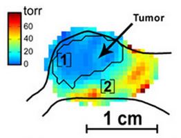
Oxygen and cancer treatment outcomeRegions of tumors with low oxygen partial pressure are important determinants of tumor biology and response to therapy. Electron paramagnetic resonance oxygen images provide accurate spatially resolved oxygen distributions in tumors. There is a strong correlation between expression of hypoxia related factors, efficiency of radiation cancer treatment and observed oxygen pressure in tumors. We explore oxygen in vivo dynamics and to find best corelates between oxygen related statistics and treatment outcome. |
Techniques and methodologies of oxygen in vivo imagingElectron paramagnetic resonance (EPR) can measure oxygen distribution in animals with 1 torr precision. Images with ~1mm resoluition can be obtained in 1-10 minutes. Prior to imaging the spin probe is injected into blood stream of an animal. In case of mice the tail vein is cannulated for injection. For larger animals more localized delivery is practiced. Then the animal is placed into an EPR imager that has a static magnetic field electromagnet, three dimensional gradient system, resonator and radio frequency bridge. EPR imaging uses frequencies in the range from 250 MHz to 1 GHz. These frequency ensure good penetration of magnetic field through the body. The magnetic fields required for resonance are much lower than the ones used for MRI and are about 100-400 Gauss. Continuous wave, rapid scan and pulse imaging techniques are used. The oxygen concentration is infered from spin-lattice and phase relaxation of spin probe. |
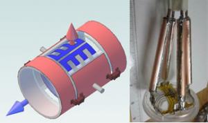
Resonator DevelopmentThe refinement of more complex aspects of biology requires higher SNR of EPR measurements. This is possible to achieve with bimodal resonators. Bimodal resonators consist of two independent resonators, which induce mutually orthogonal magnetic fields in the same volume. Using modern 3D printing techniques and electromagnetic calculation software we develop resonance structures for EPR imaging. |